Cataract and Lens Replacement Surgery
A cataract, also called "gray cataract," is a clouding of the eye's natural lens. This clouding obstructs light from entering the eye and causes patients to increasingly perceive their environment as if through fog, with poor contrast or blurriness, requiring more light for activities like reading. Additionally, increased glare sensitivity often occurs.

- Causes of Age-Related Cataracts
- The Lens Over a Lifetime
- Lens Aging
- How Common is Age-Related Cataract?
- Dr. Stefan Langenegger
- Congenital and Juvenile Cataracts
- Symptoms and Daily Life Impact
- Cataract Surgery Procedure
- Intraocular Lens Implants
- Ultrasound Method (Phacoemulsification) vs. Laser-Assisted Surgical Techniques
- Clear Lens Extraction (CLE) as an Option Before Cataract Development
- After the Cataract Surgery: Recovery and Follow-up Care
- Frequently Asked Questions About Cataract Surgery
- Further Information on Cataract Surgery
The natural lens is composed of the lens nucleus and the lens cortex. It is located within the lens capsule, which is suspended by the zonular fibers from the ciliary body.
Causes of Age-Related Cataracts
Age-related cataracts are the most common form of gray cataracts. The lens becomes cloudy due to natural aging processes - this is part of getting older and cannot be avoided.
The Lens Over a Lifetime
Over time, new lens fibers accumulate while old proteins in the lens lose their transparent structure. The lens's protective mechanisms (like certain proteins that maintain clarity) diminish over time, causing lens proteins to clump together and the lens to become cloudy.
This process occurs gradually and often goes unnoticed for a long time. In addition to losing transparency, the lens also loses elasticity and becomes thicker. The loss of elasticity leads to presbyopia (age-related farsightedness), and people who don't need distance glasses now require reading glasses.
Reduced Near Vision
By middle age, the increased rigidity of the lens becomes noticeable. Many people find that they have difficulty reading up close or working on the computer and begin to use reading glasses.
Glare
The increasing cloudiness of the lens leads to greater scattering of incoming light. This can be particularly bothersome when driving at night, as the widened pupils allow more light from oncoming cars to enter the eye.
Lens Aging
Several factors accelerate lens aging: Oxidative stress plays a major role - free radicals (aggressive oxygen molecules produced during metabolism) damage lens structures.
UV radiation from years of sun exposure increases this oxidative stress and accelerates lens aging. Diet and metabolism also influence this: a one-sided or nutrient-poor diet can deprive the lens of protective antioxidants.
General metabolic changes (e.g., in chronic diseases) can also make the lens more vulnerable. All these influences accumulate over the years and ultimately lead to age-related lens clouding.
How Common is Age-Related Cataract?
Age-related cataracts typically occur from middle to older age. Many people first notice signs of cataracts around age 60. Risk increases significantly with age: among those over 70, about every second person is affected by some degree of lens clouding.
Since almost everyone develops cataracts at a sufficiently advanced age, cataracts can be considered part of the eye's aging process. However, this isn't a sudden disease but a slowly progressive process that is highly treatable.
Congenital and Juvenile Cataracts
In rare cases, lens clouding exists from birth (congenital cataract) or develops in the first years of life (juvenile cataract). The frequency is low - about 1 in 2000 newborns has relevant lens clouding.
Nevertheless, this form is significant because it must be treated very early so that vision can develop properly in affected children.
Symptoms and Daily Life Impact
In daily life, cataracts are particularly noticeable through difficulties reading, watching TV, or recognizing faces. Problems driving are especially serious, particularly at dusk or night, as glare from headlights increases accident risk.
Cataract surgery should be considered when these symptoms significantly impair quality of life or safety - when daily activities are only possible with limitations or new glasses no longer provide sufficient vision improvement.

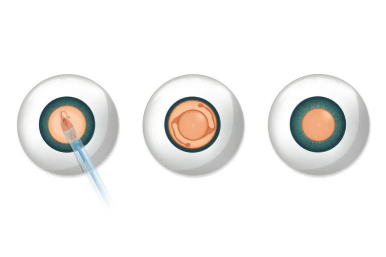
Cataract Surgery Procedure
Outpatient Surgery, Duration approximately 10-20 minutes
Cataract surgery is in most cases an outpatient procedure that lasts only a few minutes. This means you can go home the same day. A hospital stay is not necessary.
The actual cataract surgery usually takes only about 10-20 minutes per eye. Including preparation and follow-up care, you should plan approximately 2-3 hours total for the appointment.
Often one eye is operated on and the surgery on the second eye follows a few weeks later. Sometimes we decide together with you on a so-called sequential bilateral operation. In this case, the procedure is performed first on one eye and then on the second eye after a short break.
During registration, in the waiting room, and immediately before the operation, you will be repeatedly asked for your name and date of birth. This is part of the safety precaution protocol.
Anesthesia, Eye Drops and Preparation for Surgery
General anesthesia is normally not required for cataract surgery. The most common method is anesthesia with eye drops (topical anesthesia). Before the procedure, numbing eye drops are administered which make the surface of the eye insensitive to pain.
During the surgery, you may sometimes feel pressure, but no pain. Additionally, many patients receive a mild sedative so that you can lie relaxed and calm. Cataract surgery under general anesthesia is only performed in special cases for adults.
After arrival at the outpatient surgery center, your eye is prepared for the procedure with eye drops. The first drops dilate the pupil to enable problem-free access to the lens behind it. The second drops numb the eye surface so that the procedure can be performed painlessly.
Once the preparations are completed, you come into the operating room where you are already expected by your surgeon. The eye and surrounding skin are disinfected and a sterile cloth is placed over your face. Through a small nasal cannula, we supply you with additional oxygen so that you can breathe comfortably even under the cloth.
Then the surgical microscope is swung over the eye. The bright light emanating from it will usually only dazzle you at the beginning.
Now the operation begins and the natural lens is removed in several steps. During this part of the operation, you will hear various sounds as well as the computer voice of the surgical equipment.
Incision and Access
Once the anesthesia takes effect, the surgeon makes three tiny incisions at the edge of the cornea.
Fine instruments are introduced into the eye through these micro-incisions. The doctor then opens the lens capsule in a circular fashion (capsulorhexis) to access the cloudy lens.
Removal of the Cloudy Lens
The natural lens affected by cataracts is now broken up and suctioned out. This is done using ultrasound, a procedure called phacoemulsification. A fine ultrasound probe fragments the hard lens center, and the fragments are gently suctioned away. What remains is the thin, clear capsular bag that originally surrounded the lens. This capsular bag remains in the eye and serves as a "pocket" for the artificial lens.
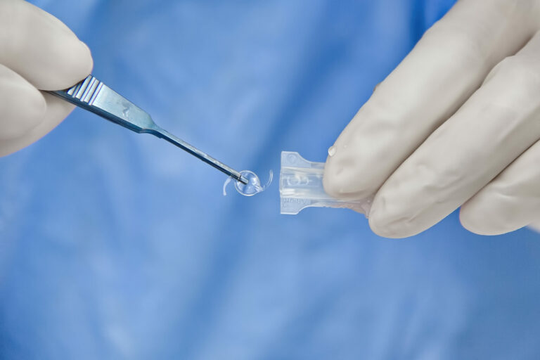
Insertion of the Artificial Lens
Now the previously selected intraocular lens (artificial lens) is implanted. It is folded so that it fits through the small corneal incision. In the eye, the lens unfolds and is positioned precisely in the capsular bag. There it remains permanently and takes over the function of the removed natural lens.
At the end of the surgery, the eye is rinsed. Usually the incision is so small that no suture is required. It closes by itself (self-sealing incision).
The actual operation is now complete. You receive a protective eye bandage and can then rest in the recovery room. Shortly afterward, you can go home, preferably accompanied by relatives.
Intraocular Lens Implants
There are a variety of different implants with various optical properties (e.g., aspherical, EDOF, multifocal, toric).
Speak with your eye surgeon about which implant and which strength (so-called target refraction) is ideal for your needs.
To prepare, ask yourself the following questions:
- How did I see in my youth? Did I need glasses and, if so, in which situations?
- In which situations would I like to be as glasses-free as possible (e.g., driving or reading)?
- What possible disadvantages am I willing to accept (e.g., certain imaging errors or contrast loss, but more freedom from glasses)?
Ultrasound Method (Phacoemulsification) vs. Laser-Assisted Surgical Techniques
As an alternative to phacoemulsification, various laser procedures have been used for many years (e.g., femtosecond laser-assisted cataract operation (FLACS) or nanolaser cataract operation). In these procedures, several steps of the operation are performed with laser assistance. In particular, the laser can be used for cutting the access points, opening the lens capsule, and actually breaking up the natural lens.
In extensive studies, the results of both methods have been investigated and compared with each other. According to current knowledge (2025), there are no reproducible advantages of the laser-assisted method. Since the costs of laser-assisted technique are much higher for patients and there is no demonstrable benefit, we do not recommend the laser-assisted method and do not offer it.
Clear Lens Extraction (CLE) as an Option Before Cataract Development
As a special case, Clear Lens Extraction (clear lens exchange) should be mentioned. This involves the same procedure as cataract surgery, but it is performed before a cataract has developed. Such refractive lens exchange can be considered, for example, in cases of high refractive error or presbyopia to enable the patient to live largely without glasses. Essentially, the still clear lens is removed and replaced with an artificial lens.
CLE is particularly interesting for people over 55 who already need strong visual aids and want to prevent future cataract surgery. We are happy to advise you whether such lens exchange makes sense in your case.
After the Cataract Surgery: Recovery and Follow-up Care
The recovery phase begins directly after cataract surgery. You remain briefly in the surgery center, and your eye is covered with a bandage or transparent eye protection. In the first 24 hours: please do not rub or press the eye and make sure that no liquids, soap, or dirt get into the eye.
Usually the first follow-up examination takes place the day after surgery. Many patients can read, watch television, or work at the computer again after one to two days, as vision improves very rapidly.
Further information about recovery and follow-up care after cataract surgery can be found here.
Frequently Asked Questions About Cataract Surgery
No. The cataract surgery itself is virtually painless thanks to anesthesia. You receive numbing drops in your eye before surgery, so you only feel pressure or touch, but no pain. If desired, you also receive a sedative to help you relax more easily.
Even after surgery, pain is rare - a slight foreign body sensation or scratching in the eye on the first day is normal, but can be easily relieved with eye drops. Severe pain hardly occurs; if it does, the eye doctor should be informed immediately.
Vision often improves shortly after surgery. Many patients can see much more clearly the next day than before. Minor fluctuations or slightly blurred vision in the first days are normal, as the eye must get used to the new lens.
The total recovery time is usually several weeks: After about 4-6 weeks, the eye is completely healed and vision is stable - at this point, if necessary, new glasses can be fitted.
In daily life, however, you are usually ready for light activities like reading, watching television, or walking after just a few hours.
Please note that each eye heals individually: Keep your follow-up appointments so that your progress can be monitored.
Usually no. The vast majority of cataract surgeries are performed under local anesthesia, either with eye drops or rarely with an injection near the eye. You remain awake during the procedure but feel no pain.
General anesthesia is only rarely necessary, e.g., in cases of great anxiety, restlessness, or certain medical circumstances. Local anesthesia is gentler on the body and carries fewer risks than general anesthesia. Your advantage: You can go home shortly after the procedure and recover more quickly.
Yes, basic insurance covers the costs of standard cataract surgery completely. This means the surgical costs, the costs for a monofocal artificial lens, as well as follow-up examinations are paid by health insurance (minus your annual deductible/franchise).
Additional costs only arise if you have special requests - for example, a multifocal lens - because these are not covered by insurance.
We discuss all such additional costs with you before surgery. So you know in advance exactly what will be covered and what you would have to pay yourself.
A true cataract does not return after surgery, because the cloudy lens was removed and replaced with a clear artificial lens - this cannot become cloudy. However, there is the so-called posterior capsule opacification: This is a clouding of the remaining lens capsule that can occur in some patients months or years later.
Posterior capsule opacification is not a new cataract, but a lighter clouding of the "skin" behind the artificial lens. If your vision becomes worse again after initially clear sight, posterior capsule opacification may be present.
The treatment is simple: The posterior capsule opacification is eliminated with a laser in a short session, without incision and without renewed surgery. Afterward, vision is clear again. This laser treatment is also covered by health insurance.
Overall, one can say: The cataract itself does not return, and posterior clouding of the lens capsule is easily treatable.
Do you have further questions about cataract surgery or would you like to schedule a consultation appointment? Our team at FIRST SIGHT Eye Clinic directly at Zurich Main Station is happy to help you.
We offer you comprehensive, individual consultation - from choosing the optimal lens implant to follow-up care - so that you can go into your cataract surgery well-informed and reassured.
Look forward to clear vision: Cataract surgery is one of the most successful procedures ever and reliably returns your visual quality and joy of life.
Further Information on Cataract Surgery
After Cataract Surgery: Follow-up Care
Cataract Symptoms: Recognizing Signs
Cataract Causes: Recognizing and Preventing Risk Factors
Freedom from Glasses After Cataract Surgery: Lens Options and Follow-up Care
Lens Options for Cataract Surgery: Advantages and Disadvantages